From an article published in Frontiers in Neuroscience in Jan. 2024.
Irie K, Ohta KI, Ujihara H, Araki C, Honda K, Suzuki S, Warita K, Otabi H, Kumei H, Nakamura S, Koyano K, Miki T, Kusaka T. An enriched environment ameliorates the reduction of parvalbumin-positive interneurons in the medial prefrontal cortex caused by maternal separation early in life. Front Neurosci. 2024 Jan 16;17:1308368. doi: 10.3389/fnins.2023.1308368. PMID: 38292903; PMCID: PMC10825025.
The Backstory: Understanding the Impact of Early Adversity
Early adverse events, such as neglect or abuse, can profoundly affect a child’s developmental trajectory. These experiences can hinder the growth of essential social skills, leading to challenges in peer interaction and emotional regulation.
The research team from Kagawa University embarked on a study to explore the underlying mechanisms of these developmental disruptions and to evaluate the potential of Sensory Enrichment to mitigate these effects.
Background and Purpose
The study aimed to investigate how early adverse events, specifically maternal separation, impact the development of social skills and the brain regions associated with these skills, such as the medial prefrontal cortex (mPFC).
Importantly, the research sought to explore whether these adverse effects could be mitigated or reversed by subsequent exposure to an enriched environment.
Environmental Enrichment (EE), characterized by increased sensory, cognitive, and social stimulation, has been shown to promote recovery in brain function and behavior.
Key Findings
Recovery in the Medial Prefrontal Cortex (mPFC)
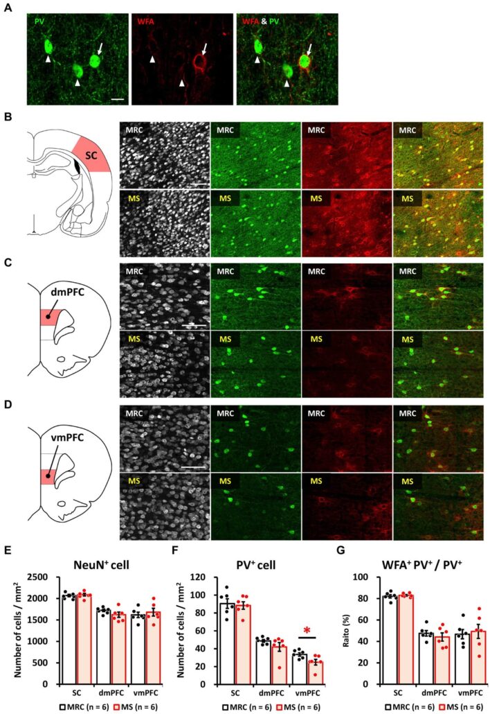
Rats exposed to maternal separation showed disturbances in the development of the mPFC, including imbalances in excitatory and inhibitory neuronal factors and a decrease in parvalbumin (PV)+ interneurons, which are critical for neural synchronization and social behavior.

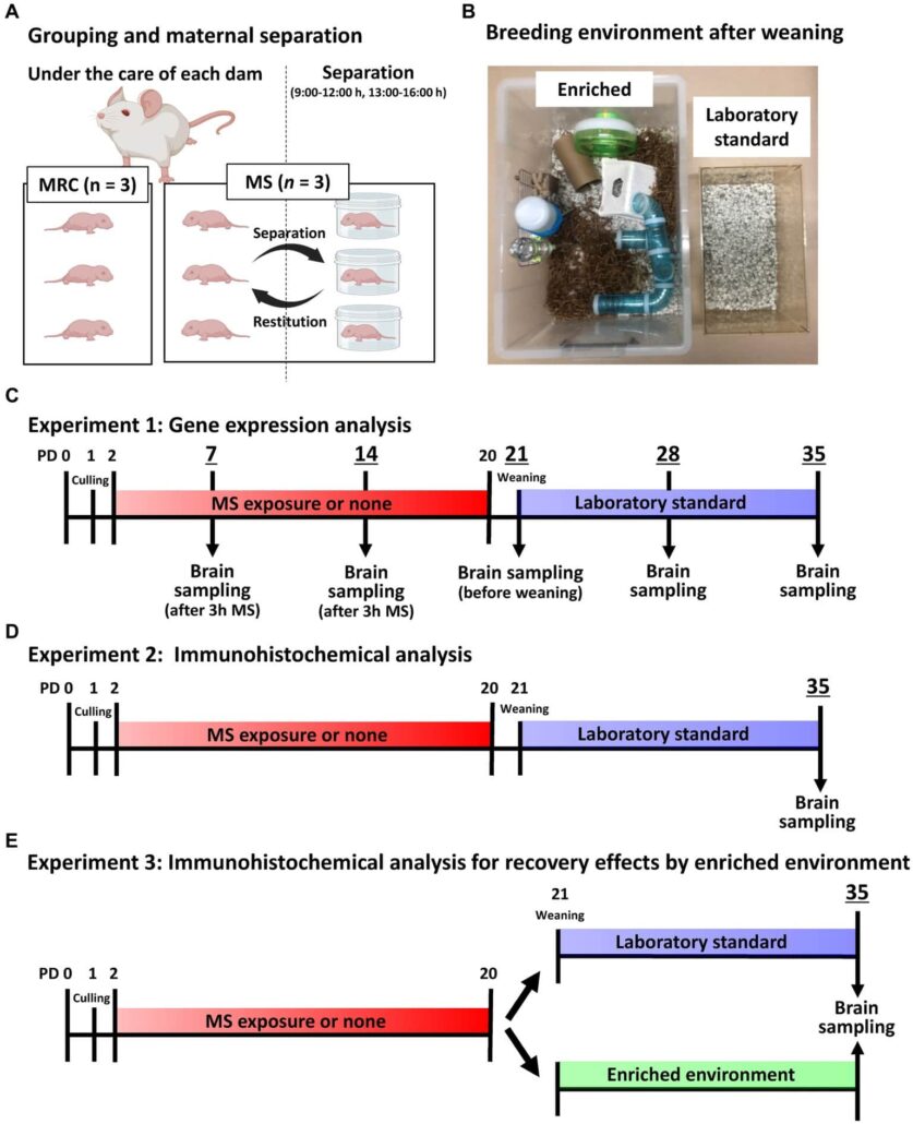
Figure 1. Image and timeline of the experimental design.
(A) An illustration of the grouping per dam and maternal separation (MS). MRC: mother-reared control. (B) Image showing the enriched and laboratory standard environments after weaning. (C) Timeline of gene expression analysis until brain sampling on each postnatal day (PD). (D) Timeline of immunohistochemical analysis on PD 35 until brain sampling. (E) Timeline of MS and enriched environment exposure for immunohistochemical analysis on PD 35 until brain sampling.
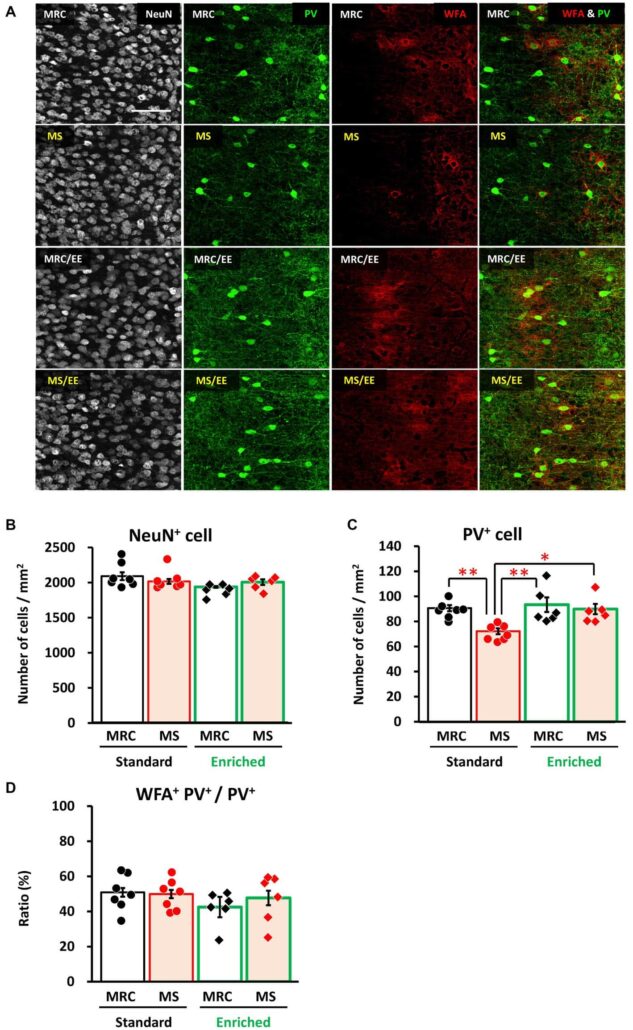
Exposure to an enriched environment after the MS period led to a recovery in the number of PV+ interneurons in the mPFC to levels comparable to those in control groups not subjected to MS.

Figure 2. Maternal separation (MS) decreased the number of parvalbumin (PV)+ interneurons in the ventromedial prefrontal cortex (vmPFC) on PD 35
(A)Immunostaining image of parvalbumin (PV)+, Wisteria floribunda agglutinin (WFA)+, and WFA+ PV+interneurons. Arrow: WFA+ PV+ interneuron. Arrowhead: PV+ interneuron without WFA. Scale bar = 20 μm. (B) From left to right, an illustration of the region of interest (ROI) for the sensory cortex (SC) and immunostaining images of NeuN, PV, and WFA, and overlapped images (PV & WFA) in both groups. Scale bar = 100 μm. (C) From left to right, an illustration of the region of interest (ROI) for the dorsomedial prefrontal cortex (dmPFC), and immunostaining images of neuronal nuclear protein (NeuN), PV, and WFA, and overlapped images (PV & WFA) in both groups. Scale bar = 100 μm. (D) From left to right, an illustration of the ROI for the ventromedial prefrontal cortex (vmPFC); immunostaining images of NeuN, PV, and WFA, and overlapped images (PV and WFA) in both groups. Scale bar = 100 μm. (E) Numbers of NeuN+ cells in each brain region in the MRC and MS groups. (F) Number of PV+cells in each brain region of both groups. (G) The ratio of WFA+ PV+ interneurons to PV+ interneurons in each brain region of both groups. The data were obtained from six animals per group and are expressed as mean ± SE. Student’s t-test or Mann–Whitney U-test were used to determine statistically significant differences between the mother-reared control (MRC) and MS groups (*p < 0.05). Contrast adjustment of images (B,C,D) was performed using identical settings based on the SC.
This suggests that the adverse effects of early stress on this crucial brain region can be at least partially reversed by environmental enrichment.
Comparison between Environmental Enrichment and Standard Environment
While the mPFC of MS rats showed persistent decreases in PV+ interneuron numbers, indicating long-term developmental disturbances, the introduction of an EE led to improvements in this area.
This recovery was not observed in rats that remained in a standard environment post-MS, highlighting the specific benefit of enriched conditions.

Figure 3. An enriched environment restored the maternal separation (MS)-induced reduction of parvalbumin (PV)+ interneurons in the ventromedial prefrontal cortex on PD 35.
(A) From left to right: immunostaining images of neuronal nuclear protein (NeuN), parvalbumin (PV), and Wisteria floribundaagglutinin (WFA), and overlapped images (PV & WFA) in each group. (B) Number of NeuN+ cells in each group. (C) Number of PV+ interneurons in each group. (D)Ratio of WFA+ PV+ to PV+ interneurons in each group. The data were obtained from seven animals per group in a standard environment and six animals per group in an enriched environment. Data are expressed as mean ± SE. After one-way or Welch’s analysis of variance (ANOVA), post-hoc tests (Tukey–Kramer or Games–Howell tests) were used to determine statistically significant differences between each group. *p < 0.05, **p < 0.01. MRC: mother-reared control. Contrast adjustment of images (A) was performed using identical settings based on the dorsomedial prefrontal cortex (dmPFC), unlike in Figure 2.
Implications for Social Behavior
Given the role of the mPFC in social behaviors, including social recognition and empathy, the recovery of PV+ interneurons in this region suggests potential improvements in these behaviors following EE exposure.
The findings support the use of environmental enrichment as a therapeutic strategy for children who have experienced early adversity.
As we reflect on the insights from this study, it becomes clear that Sensory Enrichment holds transformative potential for children who have experienced early adversities. By fostering an environment rich in sensory experiences, we can unlock the doors to improved social skills, emotional resilience, and overall well-being for these children. Mendability’s commitment to this therapeutic approach is more justified than ever, offering a beacon of hope to families navigating the challenges of developmental delays.
As we continue to explore and embrace the benefits of Sensory Enrichment, let us remember the power of our environment to shape development and healing. Together, we can support the journey of recovery for every child, ensuring a future filled with possibility, connection, and hope.

